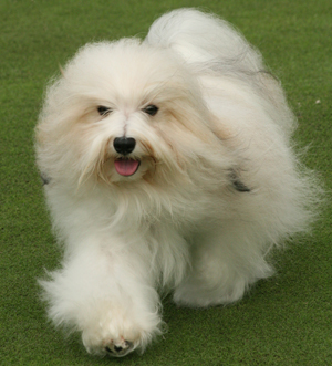
Hereditary and Genetic
The Havanese breed is relatively healthy, but there are several inherited health issues new owners should know about. They are listed below in alphabetical order.
- Cataracts
-
Cherry Eye
-
Chondrodysplasia
-
Hip Dysplasia
-
Legg Perthes (or Legg-Calve-Perthes)
-
Liver Shunt
-
Patellar Luxation (slipped kneecaps)

A web search on any of these issues will allow you to learn more about each one. To help get you started, a brief description and a few links related to each are included below.
Remember -- You are the most important part of the health care of your new Havanese. It is totally dependent on you to make sure the vet knows the symptoms and any history of health issues. Your pet is also dependent on you to ensure that preventative measures are taken, medications are given correctly and that follow-up care or testing is done. Use available resources to become more informed about potential health issues.
Cataracts
A cataract is any opacity or loss of transparency of the lens of the eye. The opacity may be confined to a small area of the lens or it may affect the whole structure. A complete cataract affecting both eyes will result in blindness. Small non-progressive cataracts will not interfere with vision. Most cataracts are inherited. There are different types of cataracts. Visit the Canine Inherited Disorders Database website for more information.
Due to the high risk of cataracts in Havanese, responsible owners and breeders should have their Hav's eyes checked (CAER eye certification) for cataracts annually. Visit the OFA eye certification website to learn more. It includes a database of canine CERF results you can search on.
Cherry Eye
A prolapse of the gland or "cherry eye" occurs when the base of the gland (embedded in the cartilage) flips up and is seen above and behind the border of the third eyelid.
The third eyelid is a triangular shaped structure in the inner corners of your dog's eyes that you may notice sometimes partly covers the eye. It consists of a t-shaped cartilage to provide support and a tear gland. The third eyelid is important in protection of the surface of the eye, and in tear production. A prolapsed gland typically becomes swollen and inflamed. Although the swelling may recede for short periods, it eventually often remains prolapsed. It is a major tear gland and should be preserved if possible.
This condition frequently occurs in both eyes and is most common in young dogs. It has not been proven that this condition is inherited, but it appears some dog breeds are predisposed. Surgery is required to anchor the gland and cartilage back into the proper position. The prolapse occasionally recurs. The gland itself must not be removed, as inadequate tear production will result causing keratoconjunctivitis sicca. Visit the Canine Inherited Disorders Database website for more information.
Chondrodysplasia
Chondrodysplasia Punctata (often referred to as CD) is the name given to a group of multisystemic, metabolic disorders of skeletal development, primarily characterized by mild to moderate growth deficiency, short stature, and bilateral or asymmetric shortening and/or bowing of the legs. Most bones in the body are first formed of cartilage, which is gradually replaced by bone early in life. Irregularities in this process will result in bones that are abnormal in size or shape. Osteochondrodysplasia describes a range of disorders such as the premature closing of growth plates, which are characterized by abnormal growth of cartilage and bone. These disorders typically result in skeletal dwarfism, with the fore limbs of the dog being disproportionately short and bowed (crooked).
Breeds such as the Dachshund and Basset Hound have been selectively bred for dwarfism. It is not part of the Havanese breed standard, but we have discovered a lot of CD in our Havs. If your Hav appears to have a 'crooked' front, your veterinarian will need to examine him to make a diagnosis. X-rays may be taken to confirm the diagnosis and to ensure there are no other abnormalities that require treatment. It is especially important to consult with your Vet if your Hav exhibits signs of lameness such as, difficulty standing or walking after getting up, decreased activity or a bunny-hop gait. Bones usually quit growing around one year old. Most cases will not require any type of surgery. However, if surgery is required, there is a better chance of recovery when it is performed while the bones are still developing.
Hip Dysplasia
Hip dysplasia literally means an abnormality in the development of the hip joint. The hip joint is a "ball and socket" joint: the "ball" (the top part of the thigh bone or femur) fits into a "socket" formed by the pelvis. If there is a loose fit between these bones and the ligaments that help to hold them together are loose, the ball may slide part way out of the socket (subluxate). Hip dysplasia can exist with or without clinical signs. When dogs exhibit clinical signs of this problem they usually are lame on one or both rear limbs. Severe arthritis can develop as a result of the malformation of the hip joint and this results in pain as the disease progresses. Many young dogs exhibit pain during or shortly after the growth period, often before arthritic changes appear to be present.
Your veterinarian may suspect hip dysplasia if your Havanese has pain or lameness in the hips. X-rays will be required to evaluate the general fit of the femur and pelvis and diagnose the problem. Usually sedation or anesthesia is required to ensure proper positioning of the dog while taking the X-rays. There are two techniques currently used to detect hip dysplasia: 1) The standard view used in Orthopedic Foundation for Animals (OFA) testing and 2) X-rays (radiographs) utilizing a device to exaggerate joint laxity developed by the University of Pennsylvania Hip Improvement Program (PennHIP). The Penn Hip radiographs appear to be a better method for judging hip dysplasia early in puppies, with one study showing good predictability for hip dysplasia in puppies exhibiting joint laxity at 4 months of age. If hip dysplasia is found, it is often possible to treat it medically or surgically.
The OFA categorizes hip dysplasia into seven different categories: Excellent, Good, Fair, Borderline, Mild, Moderate, and Severe. Visit the OFA website to learn more.
Legg-Calve-Perthes
Legg-Calve-Perthes (LCP) is another disease of the hip joints in small dog breeds. It occurs when the ball portion of the hip is damaged due to lack of blood supply. Symptoms usually appear between 5-12 months of age and involve limping, pain, and eventually arthritis. LCP can usually be confirmed with X-rays. Treatment of this condition varies according to the severity of the signs seen. Atrophy of the muscles of the affected leg is not uncommon. If atrophy is severe it can slow the recovery period considerably and may make medical therapy less likely to work. It is typically treated surgically by removing the head of the femur and letting the muscles form a "false joint." Dogs usually recuperate well from surgery. The reasons LCP occurs are not clear, however, it is assumed there may be a genetic component to the problem. Visit the Canine Inherited Disorders Database website for more information. The OFA has recently started a database for LCP.
Liver Shunts
One important function of the liver is to clear toxins, many of which are by-products of protein digestion, from the blood. A Liver Shunt occurs when a portion of blood bypasses the liver and goes directly to the heart. This allows toxins (especially ammonia) to build up in the blood stream and causes neurological signs. Symptoms include having a poor appetite, becoming lethargic, being weak or disoriented, and having seizures. It is not unusual for kidney disorders to occur in dogs with liver shunts. A special diet may keep the shunt under control. Surgery may be an option depending on where the shunt is located.
Most canines with congenital liver shunts show clinical symptoms before 6 months of age. When signs are subtle, the condition may not be diagnosed until much later. Remember, these signs can be quite vague and may include loss of appetite, depression, lethargy, weakness, poor balance, disorientation, blindness, seizures, and coma. Visit the Canine Inherited Disorders Database website to learn more.
Patellar Luxation
Patellar Luxation occurs when the kneecap (patella) pops out of joint (luxates). It is classified into several categories and grades. Signs of this problem vary based on the degree of luxation. However, it is typically associated with lameness in one of the hind legs. Visit The Orthopedic Foundation for Animals (OFA) site to learn more about it.
